Iddrisu Agbeko
CT chest without contrast
- Age
- 62 years
- Study date
- 10-09-2026
- Sex
- Male
- Accession
- 004821
Examination
CT chest without contrast
Comparison
Prior imaging: CT chest. Prior study date: 10 Mar 2026.
Technique
CT of the chest acquired in axial planes with multiplanar (coronal and sagittal) reconstructions. No acute aortic abnormality is identified on this non-contrast study.
Findings
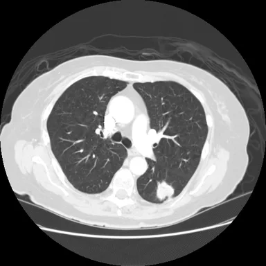
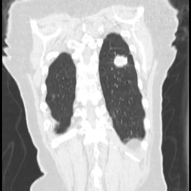
A 24 mm solid nodule in the superior segment of the left lower lobe, with lobulated margins, has grown from 16 mm on the prior study.
Lungs and airways: No other pulmonary nodule, focal consolidation or mass. The trachea and central bronchi are patent.
Pleura: No pleural thickening or pneumothorax is present.
Impression
Enlarging 24 mm solid left lower lobe nodule, previously 16 mm, suspicious for malignancy (Lung-RADS 4B). PET-CT and referral to the lung multidisciplinary team for tissue sampling.

Mawuli Dzokoto, MD
Consultant Radiologist